New Product Launch | 3D FloTrix® vivaROCK Bioreactor Stystem
2024-03-27

(Summary description)【Recap】Cellculturehasalwaysbeenthebasicandcorepartofcellbiology.Whetheritistheresearchofcellcharacteristicsorthedevelopmentofcelldrugs,itisnecessarytobaseoninvitrocellexpansiontechnology,thatis,cellcu
(Summary description)【Recap】Cellculturehasalwaysbeenthebasicandcorepartofcellbiology.Whetheritistheresearchofcellcharacteristicsorthedevelopmentofcelldrugs,itisnecessarytobaseoninvitrocellexpansiontechnology,thatis,cellcu
【Recap】
Cell culture has always been the basic and core part of cell biology. Whether it is the research of cell characteristics or the development of cell drugs, it is necessary to base on in vitro cell expansion technology, that is, cell culture technology. It is the general trend to transform the original two-dimensional cell culture into three-dimensional cell culture.
Click the picture to read the original text
For experimenters who are new to three-dimensional cell culture, how to quickly obtain an optimal three-dimensional culture process is particularly important. This article will describe the processes and key points involved in the validation phase of the 3D FloTrix® cell three-dimensional culture process.
【Preparation Stage】
1. Spinner Flask:
① The inner surface of the spinner flask is a non-attached surface. Before use, the position of the paddle should be adjusted, and the spinner flask should be cleaned, autoclaved, and dried in an oven before use.
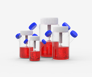
Figure: 3D FloTrix® miniSPIN Spinner Flask
② Considering the different habits and needs of users, disposable spinner flask products for bioreactors are also available in CytoNiche. The disposable spinner flasks are ready-to-use, with no need for cleaning, autoclaving and other operations.
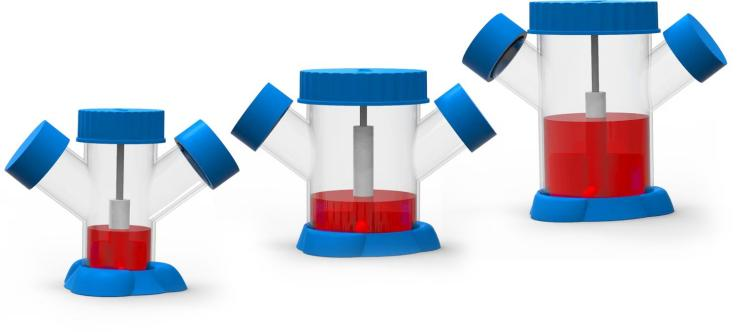
Figure: Disposable Spinner Flask
From left to right: 125 mL, 250 mL, 500 mL
Tips
① The optimal distance between the bottom of the flask and the paddle is 0.5 cm:
If the position of the paddle is too high, the liquid in the flask will be uneven. If the position of the paddle is too low, the friction between the paddle and the bottom of the flask will occur, which is not conducive to cell growth.
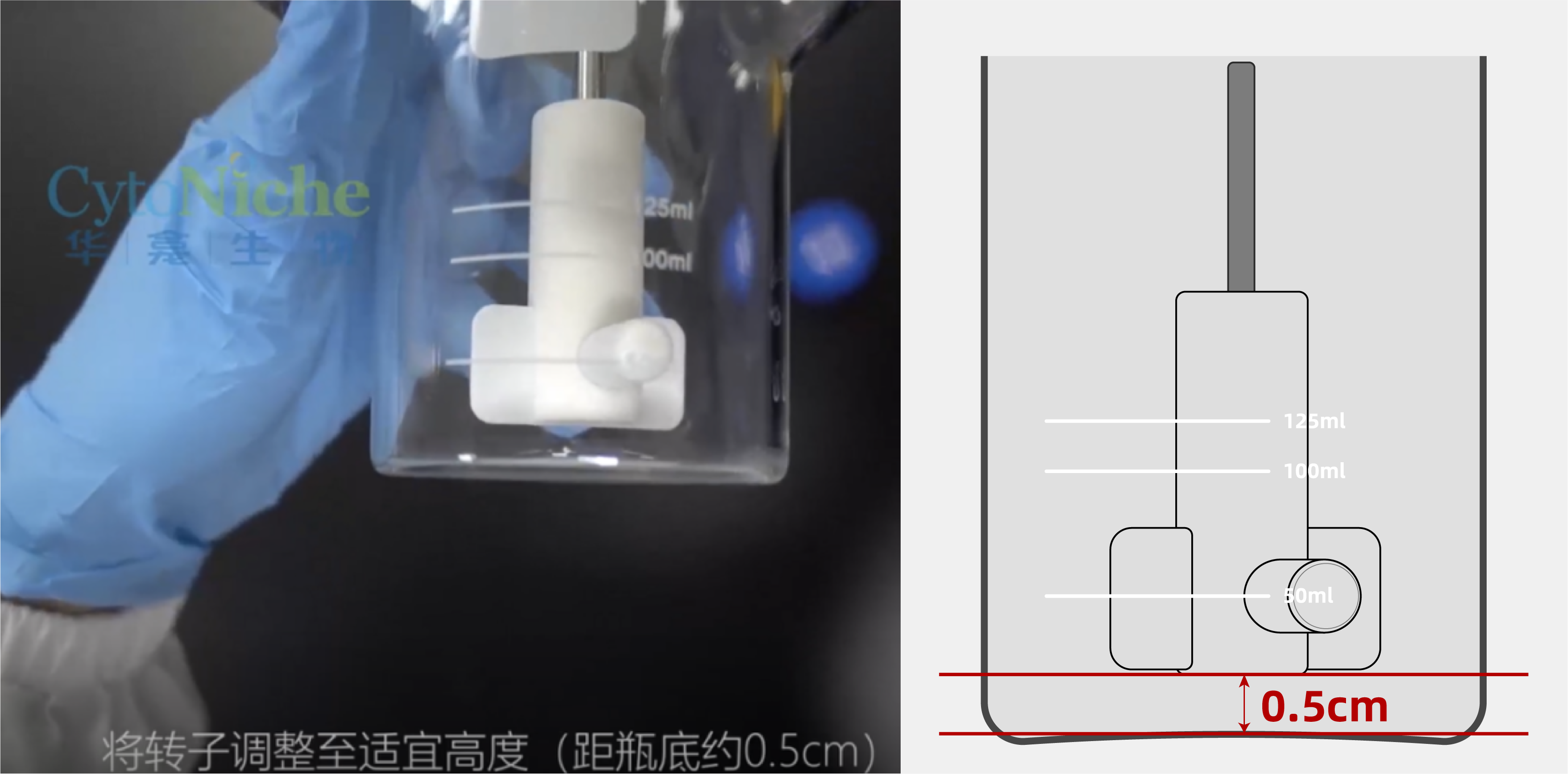
Figure: The Optimal Distance between the Paddle and the Bottom of the Flask
②Selection of spinner flask specification: Take mesenchymal stem cells as an example
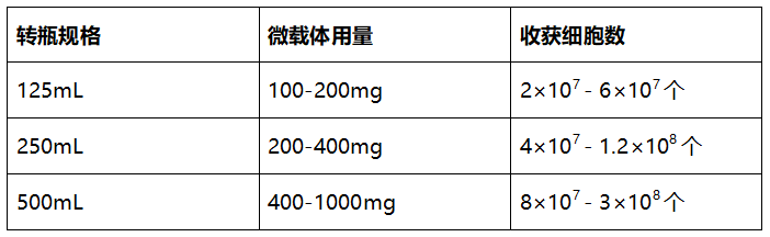
Figure: The usage of microcarriers and the number of harvested cells using spinner flasks of different specifications
2. Seed cells:
In the process of three-dimensional culture, the quality of seed cells plays a crucial role in the success of the culture.
Therefore, three-dimensional seeding requires quality inspection of seed cells, including cell doubling time, cell viability, and whether cell surface markers are qualified.
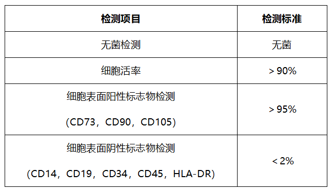
Figure: Take mesenchymal stem cells as an example
Quality Inspection Items and Criteria
Tips
① When culturing the stem cells, it is recommended to use P2-P5 cells.
② The PDT (population doubling time) of cells in two-dimensional culture is in the range of 19-20 h.
③ The digestion time of cells in two-dimensional culture should not be too long, so as to maintain high cell viability.
【2: Culture Stage】
1. Cell 3D Seeding:
Add the microcarrier tablets (ready to use) into a sterile spinner flask, add a certain amount of culture medium, evenly disperse the microcarrier tablets in the culture medium, add cells, and add enough culture medium. Place the spinner flask on the main stirring engine, set the rotation speed, and culture.
Tips
When seeding, culturing the cells with different speed can increase the cell attachment rate.
(Cell attachment rate = number of cells at 24 hours after seeding/number of cells at the time of seeding × 100%)
2. Cell culture: There are three main methods for monitoring the cell growth state during the culture process.
① Counting: After sampling, add lysate to lyse the microcarriers, count the cells, and observe the cell proliferation;
② Fluorescence staining: Use live and dead fluorescent staining solution to stain the micro-tissue (cells are bound to the carrier), and the growth state of the cells on the microcarrier can be visually observed under a fluorescence microscope. As the time of 3D cell culture increases, the number of cells on the microcarrier spheres gradually increases, as shown in the figure below.
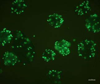
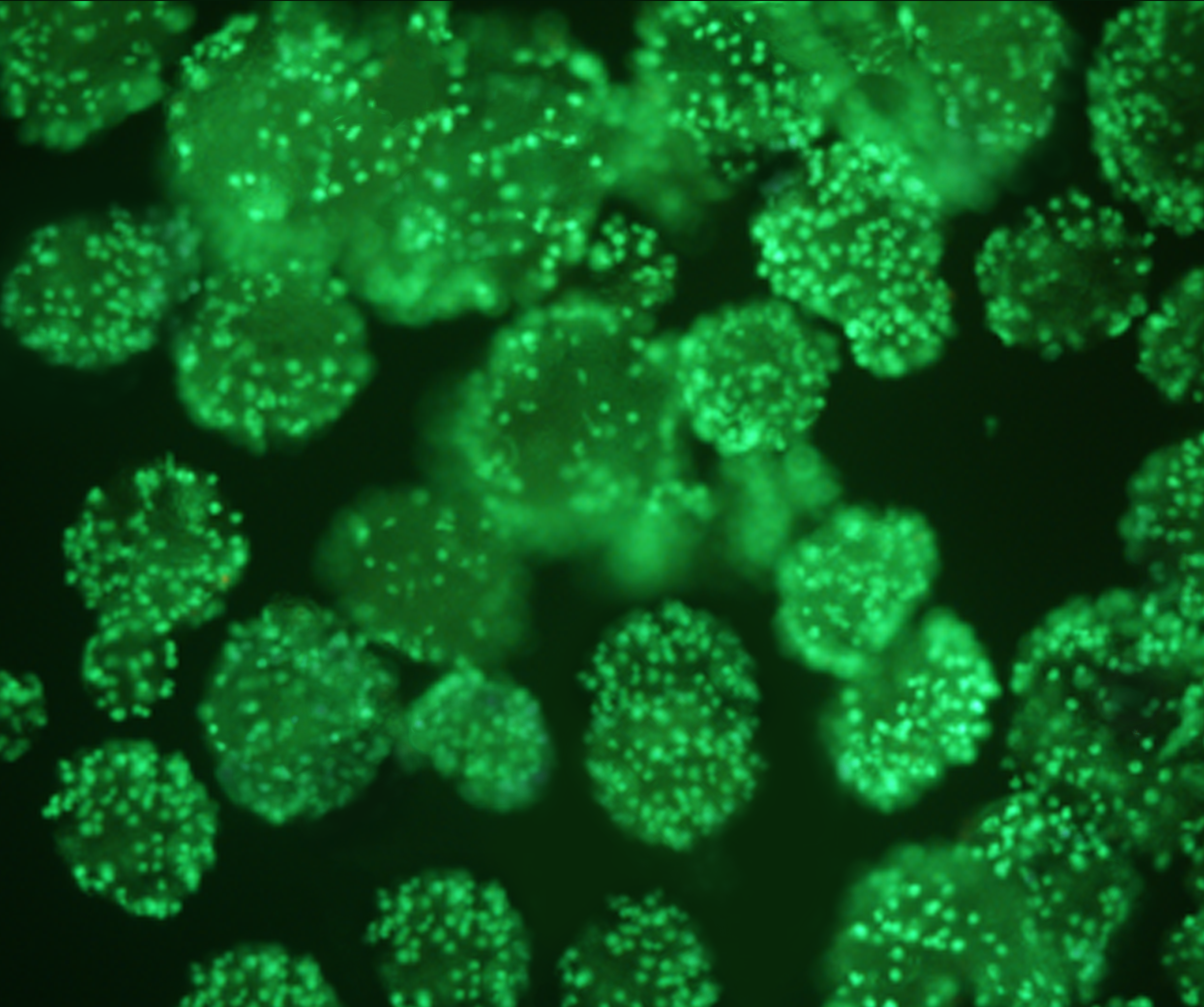
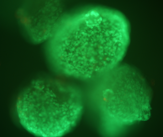
Culture for 24 h with magnification of 40x
Culture for 48 h with magnification of 40x
Culture for 96 h with magnification of 100x
③ Glucose content: Glucose in the medium will be consumed during cell culture, and the consumption of glucose will indirectly reflect the state of cell proliferation.

Figure: An example of the relationship between cell concentration and glucose content
Tips
The suitable time for lysing the microcarriers is 30 min ± 10 min.
【Three: Harvesting Stage】
When cultured in three dimensions for 4-5 days, the cells are harvested after the microcarriers are lysed with mild lysate.
1. According to the statistics of multiple experiments, using 3D TableTrix® combined with 3D FloTrix® dynamic culture process, the expansion fold of umbilical cord-derived mesenchymal stem cells (UCMSCs) is up to 10-16.
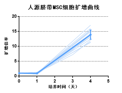
Figure: The expansion curve of human umbilical cord MSC cells
2.The UCMSC cells are continuously subcultured in 3D (the P4 cells are continuously subcultured for 6 passages), and the expansion fold of cells at 96 h could still be 12-14.
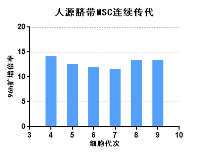
Figure: Continuous passage of human umbilical cord MSCs
【Four: Quality Inspection Stage】
The cultured cells must undergo strict cell quality inspection for subsequent applications.
1. Take mesenchymal stem cells as an example: The quality inspection is performed on three-dimensional cultured cells; the four major items are all qualified.
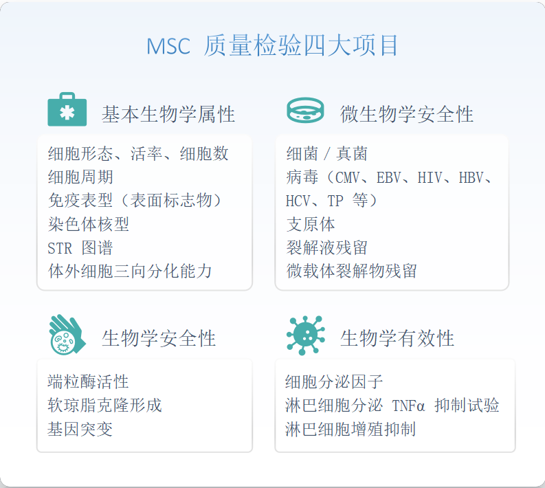
Figure: Four major items of MSC quality inspection
【3D Culture Process of CytoNiche】
As the CytoNiche 3D FloTrix® culture process is known to more and more people, an increasing number of scientific researchers are trying to use the 3D FloTrix® process for cell culture.
Many cell types are currently cultured using the 3D FloTrix® culture process. We are also looking forward to applying the 3D FloTrix® culture process to more types of cells......
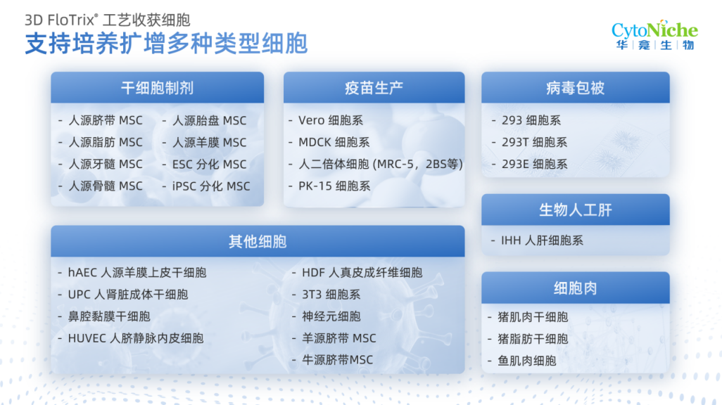
Figure: Cell types using the 3D FloTrix® culture process
【CytoNiche Technical Service Department】
The secrets are good, but our energy is limited? Don't worry~
CytoNiche Technical Service Department provides professional, refined and customized 3D cell culture services, saving you a lot of trouble and allowing you to devote your limited energy to unlimited research and development......
If you have any questions in the process of 3D cell culture, or want to quickly optimize a suitable 3D culture process, please contact us.
【CytoNiche Technical Service Department】
3D Cell Culture Field:
✦ Provide customized 3D experimental plans
✦ Customized 3D cell culture
✦ 3D cell culture process optimization
3D Cell Derivative Collection Field:
✦ 3D cell supernatant production service:
Mass production of stem cells and engineered cell culture supernatant;
✦ Exosome purification service:
Exosome purification from cell supernatants;
✦ Quality inspection service:
Detection of sterility, endotoxin, mycoplasma, protein concentration, cell secreted factors, etc. in cell supernatant;
Identification of exosomes by particle concentration, particle size, transmission electron microscopy, and immunoblotting.

Please leave your questions and needs, our technicians will communicate with you in detail
Scan the QR code to read on your phone
 京公网安备 11010802037749号
